.jpg)

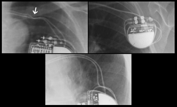
The cardiologist, cardiac surgeon, and / or nurse can later communicate with and reprogram the pulse generator with a special pacemaker programmer. The pulse generator is positioned under the skin (and sometimes also under the muscle) in the upper chest near the collar bone or in the abdominal area depending on the age and size of the patient. Leads attached to the epicardium require surgical exposure of the heart by using an incision through the chest wall. The leads are positioned within the heart with the help of a type of X-ray called fluoroscopy. Leads attached to the endocardium can be surgically placed through a vein that communicates with the heart. Leads can be attached either to the inside surface (endocardium) or outside surface (epicardium) of the heart. Single chamber pacemakers use a single lead attached to either the heart's upper chamber (atrium) or lower chamber (ventricle).ĭual chamber pacemakers use two leads, one attached to the atrium, the other to the ventricle. Pacemakers may be single or dual chamber. In addition, the leads also carry signals back from the heart to the pulse generator allowing the pulse generator to sense the heart's natural electrical activity.īy sensing the patient's natural rhythm, the pacemaker will only pace the heart when necessary. The pacing leads are flexible, insulated wires that connect to the pulse generator and carry the electronic impulse to the heart. The pulse generator is small, measuring approximately 2" x 2" x 1/4" (45mm x 45mm x 6mm) and weighing less than 2 ounces (20-30g). These circuits contain timers that regulate how often the pacemaker must send impulses to stimulate the heart. The pulse generator houses the battery and electronic circuits (like a small computer).

A pacemaker is a small, battery-powered medical device designed to electrically stimulate the heart muscle in an effort to restore the heart rhythm towards normal.Ī pacemaker system consists of two main parts: the pulse generator and pacing leads.


 0 kommentar(er)
